
Brain Morphometry Patterns of Mixed AD and LATE

Overview
Alzheimer’s disease neuropathologic change (ADNC) and limbic-predominant age-related TDP-43 encephalopathy neuropathologic change (LATE-NC) frequently co-occur in late life, but their independent vs. combined neurodegeneration patterns are still being clarified. In this project, we used voxel-wise deformation-based morphometry (DBM) on ex-vivo MRI (N=912) from a community-based autopsy cohort to map brain morphometry patterns across four pathology groups (AD± / LATE±), while controlling for other comorbid neuropathologies, demographics, and scanner.
Key findings
- AD+LATE- and AD−LATE+ each showed less tissue primarily in medial temporal lobe structures (overlapping signatures).
- Mixed AD+LATE+ showed more widespread tissue loss across temporal, frontal, and parietal regions-consistent with additive/synergistic neurodegeneration.
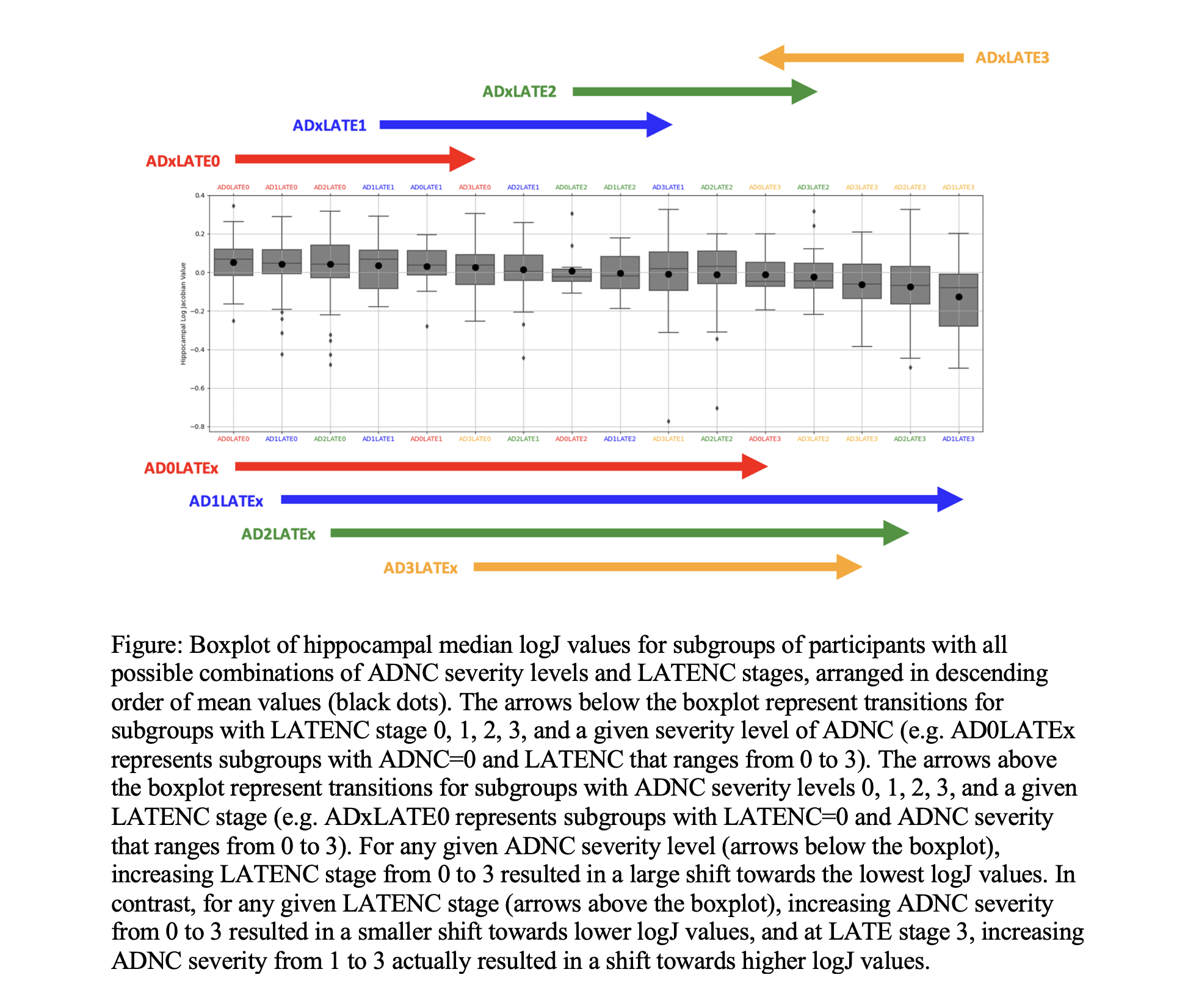
- LATE+ was associated with a smaller anterior hippocampus compared to AD+ alone, and LATE stage increases showed a stronger relationship with hippocampal shrinkage than ADNC severity increases.
- Patterns were also present but less pronounced in participants without dementia.
Resources
ISMRM 2024 · Abstract · Watch my talk in ISMRM 2024
AAIC 2024 · Abstract