
Neurofibrillary Tangles Prediction Based on MRI
Date:
Slides: Download
Neurofibrillary tangles are a hallmark of Alzheimer’s pathology and are closely linked to brain atrophy and cognitive decline. In this talk, I presented an MRI-based classifier for tangles that leverages ex-vivo MRI + autopsy-confirmed neuropathology from a large community-based cohort.
Approach
- Cohort & imaging: 878 autopsied older adults from Rush longitudinal cohort studies (MAP/ROS/MARS) with ex-vivo 3T MRI and detailed neuropathologic examination.
- Model: L2-regularized SVM to classify Braak V–VI vs. 0–IV, using demographics (age, sex) plus multimodal MRI-derived features (volumetry, cortical thickness, subcortical shape, diffusion, R2).
- Evaluation subset: performance reported on the subset with all feature groups available (N=74).
Key result
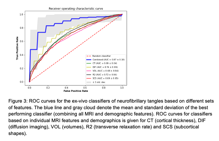
The combined model achieved AUC = 0.87 with 82% sensitivity and 77% specificity, outperforming a published in-vivo MRI+pathology approach reported at AUC = 0.69.
Why it matters
Successful ex-vivo → in-vivo translation of this work could enable a non-invasive tangles classifier to support refined participant selection and targeted therapies.
Photos and Figures from the Conference
Figure 3 - Classifier performance (ROC)
Conference photo

Group photo from the AAIC 2022 lightning session (including the moderator Dr. Lea Grinberg, M.D., Ph.D.).