
Subcortical Volume & Shape Signatures
Overview
Deep gray matter structures are impacted early in aging-related neuropathologies, but these pathologies often co-occur, making it hard to isolate their independent effects. In this project, we combined autopsy-confirmed neuropathology with ex-vivo MRI to quantify volume loss and localized shape deformation across key subcortical structures in a large community-based cohort.
What I did
- Performed multi-structure segmentation and volumetry on ex-vivo MRI.
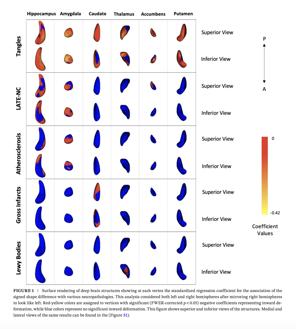
- Conducted vertex-wise shape analysis using SPHARM-PDM to map localized inward deformation patterns.
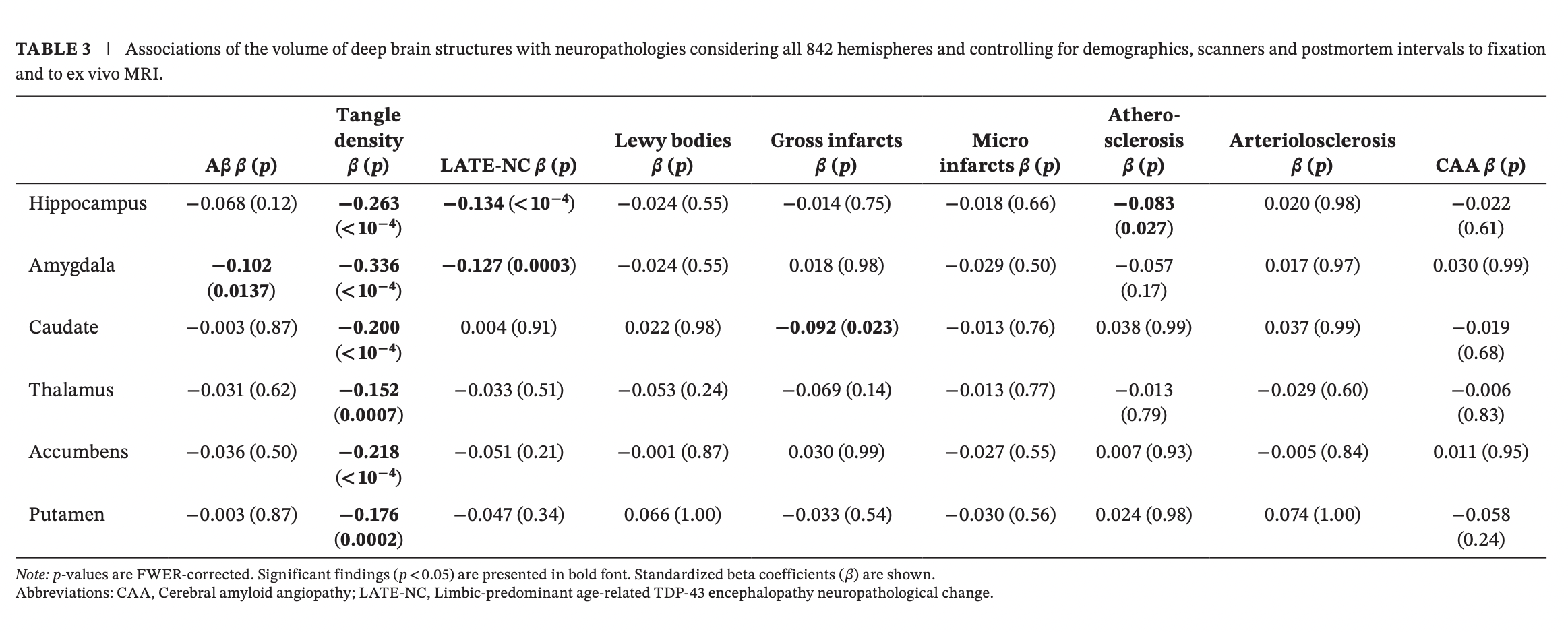
- Ran permutation-based regression (PALM) to estimate independent associations of multiple neuropathologies with structure-specific volume and shape metrics, controlling for demographics, scanner, and postmortem intervals.
Key findings
- Tangles showed broad, independent associations with structural abnormalities across all deep gray matter structures.
- LATE-NC was independently associated with abnormalities in the hippocampus and amygdala (with additional localized shape effects in nucleus accumbens).
- Atherosclerosis showed independent associations with hippocampal abnormalities.
- Gross infarcts showed independent associations with the caudate.
- Shape analysis revealed distinct spatial “signatures” that help disentangle mixed pathologies beyond what volume alone captures.
- Volume and shape analyses converged on the same core pathology–structure associations, while shape analysis added spatial “deformation signatures” and revealed additional pathology links beyond what volume alone captured.
Resources
Human Brain Mapping 2025 · Paper
ISMRM 2023 · Abstract
AAIC 2023 · Abstract